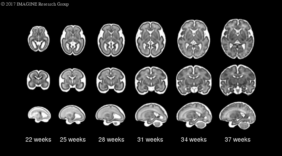
The fetal brain undergoes rapid changes during the second and third trimesters of gestation. In addition to overall growth, the brain dramatically changes in terms of shape, with the gyrification or folding of the cerebral cortex, and also in characteristic, as layers of neurons become myelinated. At the same time, the challenges of imaging in-vivo neonates, such as fetal and maternal motion, limited resolution, and image artifacts result in corrupted images or low-quality data. The lack of quality MR images throughout this time period and the need for improved data processing techniques for challenging datasets necessitate improved digital reference images or brain atlases.
To this end we have developed a normative spatiotemporal MRI atlas of the fetal brain. The atlas includes 3D (spatio-) images of the fetal brain in isolation at a range of gestational ages (-temporal). The atlas was constructed from 81 fetuses in normative pregnancies, combined using symmetric diffeomorphoic deformable registration in space, with the influence of contributing cases weighted by age. The resulting atlas images are high-resolution representations of the average developing fetal brain at one-week intervals between 21 and 38 weeks gestational age and can be used for anatomical reference and measurement.
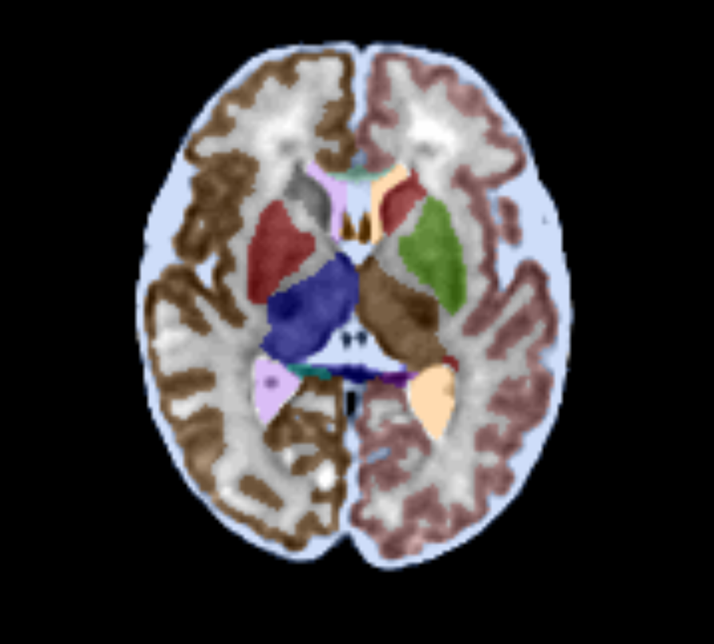
We have segmented the atlas images facilitating the measurement and localization of individual structures (i.e. hippocampus, corpus callosum), tissue classes (i.e. white and gray matter, cerebrospinal fluid), and developmental white matter layers (i.e. subplate) in the brain. These segmentations can be used to delineate novel fetal brain images through the use of algorithmic registration and segmentation programs that use the atlas images for reference. They can also be used to provide information such as the thickness of cortical plate regions or to designate the location of neuronal tracts measured by diffusion-weighted imaging.
You can download the fetal atlas or check it out in a browser-based interactive 4D viewer.
Publications
- A Gholipour, CK Rollins, C Velasco-Annis, A Ouaalam, A Akhondi-Asl, O Afacan, C Ortinau, S Clancy, C Limperopoulos, E Yang, JA Estroff, and SK Warfield. A normative spatiotemporal MRI atlas of the fetal brain for automatic segmentation and analysis of early brain growth, Scientific Reports 7, Article number: 476 (2017).
- A Gholipour, C Limperopoulos, S Clancy, C Clouchoux, A Akhondi-Asl, J A Estroff, and S K Warfield. Construction of a Deformable Spatiotemporal MRI Atlas of the Fetal Brain: Evaluation of Similarity Metrics and Deformation Models. MICCAI 2014.