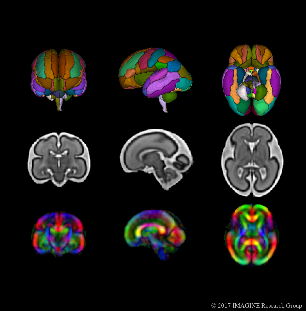
Diffusion-Tensor Imaging (DTI) is an integral aspect of brain imaging with MRI technology. Examining the diffusivity or movement of water molecules allows us to glean information about the structural pathways, connectivity, and underlying patterns of brain developement- and variation in the prescence of congenital disorders- that cannot be characterized solely using structural imaging. The application of DTI techniques to in-utero fetal imaging, however, remains challenging. Like in structural imaging, motion (both fetal and maternal), signal-to-noise ratio (SNR), and inhomogeneity artifacts remain major obstacles preventing the consistent acquisition of high-quality, fetal DTI data, and thus the development of reference atlases and automated analysis methods.
To address these issues, we have developed a robust computational framework for the construction of a in-utero, fetal DTI atlas (available through the link below). The atlas was constructed from tensor maps of 67 in-utero DTI scans of fetuses in normative pregnancies. The results of the atlas construction were validated through comparison with previous in-utero and pre-term infant DTI studies, and ex-vivo DTI and histological studies. The atlas release includes fractional anisotropy (FA), color fractional anisotropy (CFA), and Mean Diffusivity (MD) maps for gestational age weeks 22 through 38.
The download link for both the structural and DTI fetal atlases can be found here.
Publications
- Khan S, Vasung L, Marami B, Rollins CK, Afacan O, Ortinau CM, Yang E, Warfield SK, Gholipour A. Fetal brain growth portrayed by a spatiotemporal diffusion tensor MRI atlas computed from in utero images. NeuroImage. 2018 Aug 30.
- Khan S, Rollins CK, Ortinau C, Afacan O, Warfield SK, Gholipour A. Tract-Specific Group Analysis in Fetal Cohorts using in utero Diffusion Tensor Imaging. MICCAI 2018 (upcoming)